Scalp injuries- Explained by Dr Saad Andalib ( A review article).
Scalp hematomas:
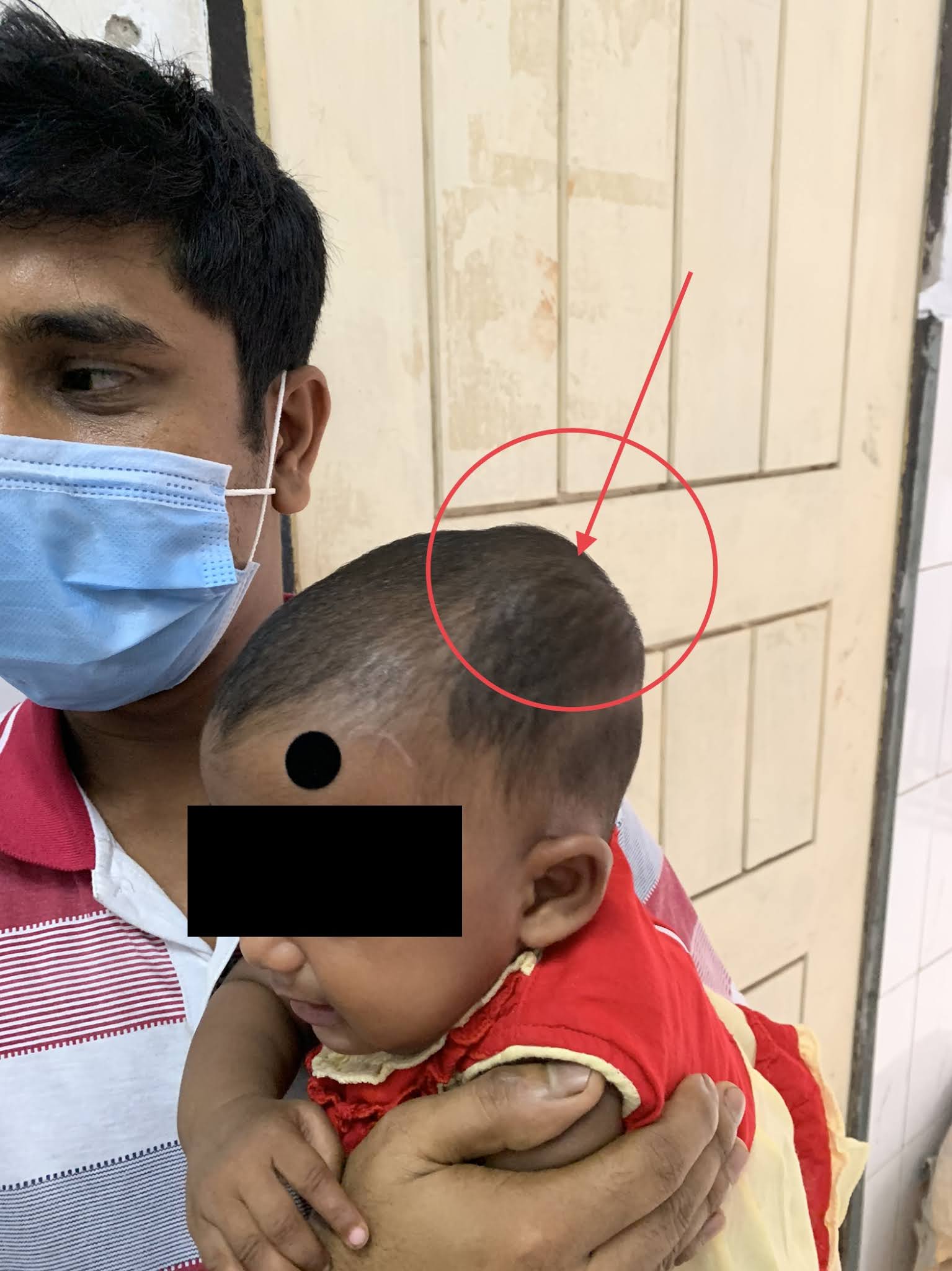
Scalp hematomas are an important indicator of potential TBI, especially when they appear in younger infants (eg, <6 months of age), are larger (eg, >3 cm), and are located in the temporal, parietal, or occipital regions.


Hematomas of the neonates:
Introduction:
Neonatal hematomas refer to a grouping of extracranial injuries that occur during delivery and are secondary to edema or bleeding into the varying locations within the scalp and skull.

Caput Succedaneum:

- Edematous region above the periosteum that crosses suture lines
- Presents at birth, typically after prolonged or difficult labor due to compression against bony prominence of maternal pelvis
- Visualize pitting edema on physical exam
- Discoloration may be present
- Usually resolves within a few days and requires no further treatment
- Complications to look out for include long term scarring and alopecia
- Halo scalp ring is an alopecic ring that can develop after resolution.
Cephalohematoma:

- Subperiosteal bleed due to rupture of vessels beneath the periosteum
- Presents right after delivery as swelling that does NOT cross suture lines
- Can have some discoloration
- More common when forceps or vacuum delivery is performed
- Usually doesn’t expand after delivery
- If one notices expansion pursue imaging and work up for source of continuing bleed
- Resolves spontaneously over course of a few weeks
- May cause indirect hyperbilirubinemia due to absorption of the bleed
- Monitor for calcification and ossification, which can result in deformity of skull
- If the cephalohematoma becomes erythematous and fluctuant, infection might be present
- Most commonly due to E. coli infection
- Must do incision and drainage of abscess and debridement of necrotic skull if needed.
Subgaleal hematoma:

- Bleed located between periosteum of skull and the aponeurosis
- Presents as fluctuant swelling of the head that may shift with movement
- Rapid loss of intravascular volume causes tachycardia and pallor
- Potential for loss of 20-40 percent of neonate’s blood volume
- Most are due to vacuum-assisted delivery, so monitor for following those deliveries
- Develops around 12-72 hours after delivery
- Early recognition is most important for survival
- Once suspected, monitor by serial measurements of hematocrit and frontal circumference
- Volume resuscitation with packed red blood cells, fresh frozen plasma, and normal saline to stabilize vitals
- May need surgical evacuation.
References
- Chang, H. Y., Peng, C. C., Kao, H. A., Hsu, C. H., Hung, H. Y., & Chang, J. H. (2007). Neonatal subgaleal hemorrhage: clinical presentation, treatment, and predictors of poor prognosis. Pediatrics International, 49(6), 903-907.
- Ferriero, D. M. (2004). Neonatal brain injury. New England Journal of Medicine,351(19), 1985-1995.
- McKee, T. M. (2015). Neonatal birth injuries. Uptodate.
- Nicholson, L. (2007). Caput Succedaneum and Cephalohematoma: The Cs that Leave Bumps on the Head. Neonatal Network, 26(5), 277-281.


Comments
Post a Comment